


Ankle Dorsiflexion is an Often Overlooked Cause of Pain Throughout the Body. Working on Dorsiflexion Range of Motion will Aid Your Knees, Hips, Pelvic Floor, Low Back, Neck, and Even Your TMJ and Jaw.

Updated: 9 hours ago
What is Foot Dorsiflexion?
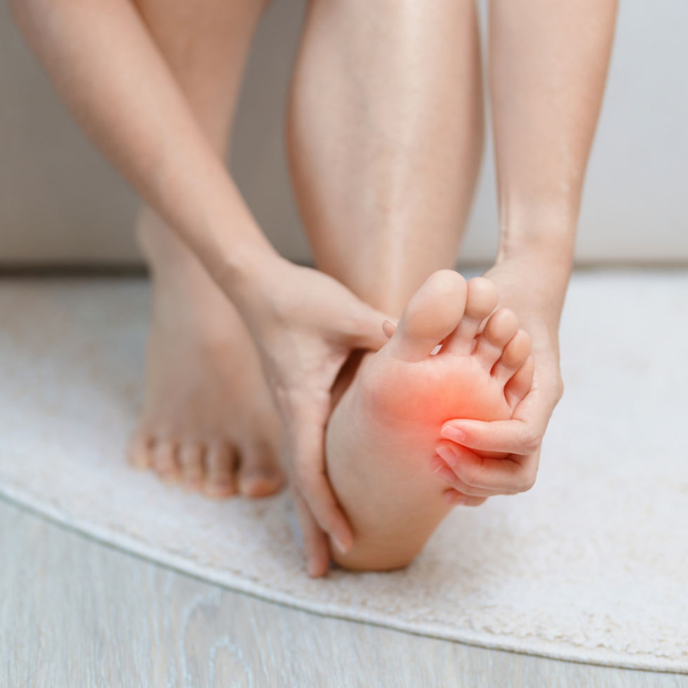
Dorsiflexion can refer to hand or foot (and even toe) movement. This post will focus on foot dorsiflexion. The dynamic ankle joint is called the talocrural joint and serves foot motion in two fundamental ways: plantar flexion and dorsiflexion. Plantar flexion is when the toes point downward, and dorsiflexion is when the toes point upward. Dorsiflexion of the ankle joint is happening as the toes come closer to the shin. Dorsiflexion is happening when you are walking, running, jumping, and doing a squat.
During a squat, with your feet planted firmly on the ground, dorsiflexion happens as your knees move over your toes. If you lack ankle dorsiflexion, you will unknowingly compensate by leaning forward with your torso to complete the squat. When the body compensates for dorsiflexion, a cascade of other dysfunctional compensations will travel all the way up the body.
What You Will Find in This Article on Dorsiflexion
The really "fun" section of this post is called “Healthy Dorsiflexion is Essential For…..EVERYTHING!” and in that section you will find a list of things that dorsiflexion affects such as ACL injuries and the pelvic floor! Skip ahead and scroll to the pink arrow to find that fascinating section.
This post will also cover common issues that hinder dorsiflexion and list muscles that create imbalance. If you want to see a longer list of lower muscles and their function head to a blog post HERE
This post will also briefly speak to shoes and dorsiflexion and give you an idea as to how to test your own dorsiflexion at home.
A lack of dorsiflexion can create an anterior pelvic tilt and internal hip rotation. I have other articles COMING SOON!

Your Body is Connected
I am writing this blog post to aid women I see in my practice who have jaw and neck pain, and diaphragm and pelvic floor pain and dysfunction. In my practice, I spend an hour at a time helping women remove soft tissue restrictions and lighting those areas of the brain so the mind-body can create its own change.
Important: Often when I am working on the jaw, women say they feel changes happening all the way down to their feet. When I am working on the pelvic floor, and a woman is feeling anxious, gripping, or holding onto tension, I will instead spend time inside the jaw. When we return to the pelvic floor—like beautiful magic—the holding pattern in her pelvic floor will have changed, softness becoming more possible. When we work with the diaphragm, corrections in the ribcage and pelvic floor become possible, and then corrections in the neck and jaw are more profound.
I do not work with dorsiflexion in the office (but will be teaching foot classes sometime this year!) This article is a resource as dorsiflexion is affecting the rest of the body; the things I work directly on in the office.
All of your body is affecting all of your body, and that can sometimes feel overwhelming. But through education, choosing to work on a few things at a time, developing a health-providing fitness routine, realizing you have individual needs, and realizing that perfection is not necessary, you can begin the journey of connecting to and understanding—with gentleness and compassion—the needs of your mind-body system.
What Hinders Dorsiflexion?
Like all joint movements, dorsiflexion can become hindered. If any joint in your body feels hindered, it suddenly feels like the most important one :) However, when the foot’s dorsiflexion is hindered, your entire body—from the foot upwards—will be under that effect. Researchers have shown that dorsiflexion lights up many areas of the brain.
Using the highly innervated and sensitive sole of your foot, how your foot perceives the ground and how your foot navigates each step, your brain is taking in information and making quicker-than-lightning decisions.
Dorsiflexion can be hindered for several reasons; soft tissue restrictions, muscle imbalances, and joint inflammation. This blog post will use the abbreviation ROM when discussing a lack of range of dorsiflexion motion. ROM means “range of motion."
A quick explanation of how joints and muscles work together: Muscles move joints in the desired direction(s) and other muscles work against those motions. These opposing forces support joint health.
Muscles that work to create a joint’s motion are called agonist muscles. Muscles that work against the motion are called antagonists. Antagonist muscles create stability.
But when there is an imbalance, when antagonist muscles are hypertonic—tight and tense—or have adhesions and fascia restrictions, the antagonist muscles create restriction rather than stability. And when under weight load, the agonist muscles, the muscles that produce the joint motion, may be too weak to produce a motion to the joint’s full capacity. This muscle imbalance can cause local pain, but also causes compensation patterns to develop elsewhere in the body.
Again, if it helps you, I have a blog post HERE that discusses each of these muscles origins, insertions, and actions. I also have a page on my website that has links to shoes and some other recommended foot health products (including spikey mats--my favorite!) in my shop HERE Check out my other shop pages as it is all stuff I recommend in the office.
Scroll Down to the PINK ARROW for the List of What Dorsiflexion Affects!
Muscles and Joint Capsule Adhesions
Gastrocnemius and Soleus
Muscles on the back of the calf, the gastrocnemius and soleus, are antagonists to dorsiflexion. Dorsiflexion is when the foot moves toward the shin, and the gastrocnemius and soleus are on the back of the calf. When these muscles are overly tight and tense, they restrict dorsiflexion.
The Achilles Tendon and Plantar Fascia
The achilles tendon and plantar fascia can also restrict joint movement.

Tibialis Anterior, Extensor Digitorum Longus, Extensor Hallucis Longus, Fibularis Tertius
The muscles that produce the action of dorsiflexion are the tibialis anterior (a major dorsiflexor), extensor digitorum longus, extensor hallucis longus, fibularis tertius (also called peroneus tertius-how confusing!)
Weakness of these primary mover muscles will hinders dorsiflexion. Weakness in these muscles can happen progressively after motion is restricted. By that I mean that if there is a restriction caused by antagonist muscles, and that restricts joint motion, the muscles that create the motion will fall into disuse. When they are not being used, they will become weak. This is one of the fundamental principles of manual massage therapy and why manual massage therapy is so effective. Manual therapy aims to reduce restrictions, which allow muscles to have greater ROM and natural strength.
Joint Capsule
Fibrous adhesions in the joint can also cause a lack of proper joint movement, and the ankle—like the shoulder—can even experience adhesive capsulitis (a “frozen” joint). If when testing your ankle’s dorsiflexion mobility you feel a pinch at the front of the ankle, you likely have a restriction in the ankle joint itself—opposed to soft tissue restrictions. But again, both joint issues and soft tissue restrictions can be paired together, and soft tissue restrictions will eventually create joint issues.
Bony Impairment
A bony impairment—a bone spur—can also hinder motion.
Different Types of Ankle Movement
A lack of range is also called hypomobility. Excessive motion or hypermobility of the ankle joint also exists. In this recipe, we are talking about hypo mobility. Hypermobility of the ankles causes by genetic factors, such as inherit ligament laxity that happens in Hypermobility Spectrum Disorder and Ehlers-Danos. Injury and weakness of the muscles around the ankle can also contribute to hypomobility.
What is Healthy Dorsiflexion?
Literature stating the ideal range of motion for ankle dorsiflexion varies. And unless you are working with a professional who is helping you measure, I do not suggest you worry about what range of motion you have in terms of degrees. But I do suggest you consider dorsiflexion in terms of availability of functional movement. At the end of this blog post, I will outline a way that you can asses your dorsiflexion at home.
You can also focus on how you feel during daily activities and fitness, and note what the signs of ankle dysfunction are—listed below. If you are used to a lack of range of motion, you might not realize it. But in those cases, checking in with calf tension, or the ability to perform a comfortable squat, or walk uphill, will help you realize that dorsiflexion should be improved.
Normal range of motion for a non-weight bearing ankle is 10-20 degrees. Walking on a flat surface requires around 10 degrees of dorsiflexion. Dynamic motion such as running or squatting requires up to 20 degrees of dorsiflexion and may even require up to 30 degrees for running or sprinting.
You can still perform dynamic activities with less than an ideal range of motion, but you will then compensate with other muscles and joints, and that will cause structural issues distal (away from) the ankle joint.
You may suffer from a lack of dorsiflexion, but it will be another joint that expresses an issue.
Many types of practitioners can assess ankle dorsiflexion, but again, if you have recurring knee or hip issues, you can check in—on your own—with dorsiflexion. And of course, there is always You Tube and people on that platform will be able to demonstrate dorsiflexion for you! Squat University is a great resource for any woman who partakes in squats or functional fitness. One of the things that a lack of dorsiflexion creates is tibial rotation. I love this article by Squat University that describes leg position and motion, cause and effect, of tibial rotation during squats: https://squatuniversity.com/2023/04/12/what-is-tibial-rotation-why-you-should-know-about-it/
And whereas I normally list all resources at the end of a post, here is a link to one of the best articles I have read on dorsiflexion (although I have simplified much of the important information in this post): https://www.ejgm.co.uk/download/role-of-ankle-dorsiflexion-in-sports-performance-and-injury-risk-a-narrative-review-13412.pdf
Here is the "fun" part of this article ;) Learn all the things that dorsiflexion affects.

Healthy Dorsiflexion is Essential for…..EVERYTHING! Learn what dorsiflexion affects in the rest of the body HERE:
When ankle dorsiflexion is less than 10 percent, the gait cycle alters. The gait cycle is the sequence of movements that occurs during walking. A limited range of motion in the ankle joint will create short steps. Knee and hip function will become altered.
The changes that altered dorsiflexion create in the body are fascinating! Here is a list of the varied changes that occur when you alter dorsiflexion, and therefore alter your gait cycle and the way you move during sports and fitness:
Between the two ankles, there can be asymmetry in dorsiflexion and therefore asymmetry in the gait cycle and asymmetry with knee and hip movement. In a study on army rangers, those with asymmetry measuring at 6 degrees were 5.1 times more likely to suffer other forms of musculoskeletal overuse injuries.
Proper dorsiflexion actually recruits your transverse abdominal muscles. Proper recruitment of your transverse abdominal muscles aids in maintaining posture, aiding each part of your body. Transverse abdominal muscles improve balance and overall stability, and you need them to engage in fitness and exercise healthily.

Decreased dorsiflexion creates decreased transverse abdominal muscles, and decreased transverse abdominal activity is associated with increased low back pain. And studies have shown that those with mechanical issues in the low back, resulting in low back pain, also have decreased dorsiflexion ROM.
Decreased ROM in dorsiflexion will lead to altered movement patterns in the other joints.
Those with limited ROM in ankle dorsiflexion experience increased adductor activation compared to their gluteal muscles. As a pelvic floor therapist, I would add to this is that adductor tension creates pelvic floor strain (post on adductors, anterior pelvic tilt and pelvic floor health COMING SOON).

In women, healthy dorsiflexion allows for a greater and healthier contraction of the pelvic floor muscles. VERY interesting!

A lack of dorsiflexion increases your risk of hamstring injuries.
When in a fitness class, you will move your body from side to side. When talking about these motions in terms of anatomy, we would say these are frontal plane motions. An example of this would be lateral lunges and resistance band side steps. Decreased ankle ROM will cause your body to compensate during frontal plane motions.

Gastrocnemius and soleus hypertonicity and stiffness lessen knee flexion, while producing greater hip adduction (the leg moves closer to the midline of the body), and knee external rotation. Over time, a lack of range of motion in flexion and extension will create overload knee injuries and pain in the front of the knee. A lack of flexibility in the gastric and soleus will affect the knee during motions such as lunges and squats.
During functional activities, altered gastrocnemius will change knee function and further Achilles tendon strain. Ground force strain to the Achilles tendon will lead to a 3.6 times greater chance of developing painful tendinopathy.
Pronation is your foot landing on the foots outer edge. Neutral pronation helps your weight to disrupt evenly when you walk. A lack of dorsiflexion can cause overpronation and your weight will not be evenly distributed.

Overpronation will cause you to loose the arch in your foot leading to flat feet and furthering increasing your risk of injury. Developing a flat foot, loosing the arch, will further increase overpronation.
Overpronation will increase femur and hip internal rotation which overloads the knee.
Dorsiflexion ROM problems will increase the risk of developing plantar fasciitis. Reduced range of motion in the gastrocnemius and a lack of dorsiflexion flatten the arch of the foot. This causes increased pressure during the stance phase of walking, which comprises 60 percent of the gait cycle. This can lead to ligamentous structures being overstretched and can cause inflammation in the plantar fascia.

Decreased dorsiflexion will also cause you to compensate when jumping and squatting. Decreased dorsiflexion during squats will cause internal rotation of the knee(s). Decreased dorsifelxion in one or both feet will create knee valgus (also called knock-knees).
A lack of dorsiflexion ROM will also cause the quadriceps to show reduced activation during squats while the soleus will demonstrate enhanced activation. As you will recall, hypertension (holding too much tone and tension) in the soleus muscle causes a lack of dorsiflexion. This shows a cycle.
A lack of dorsiflexion has been linked to ACL ruptures and other functional overload injuries like patellar instability and pain around the iliotibial band.

A lack of dorsiflexion will alter the relationship between the tibia and the femur. The ACL plays a vital role in stabilizing that joint during movement. Whereas the quadriceps can play a role in a “dysfunctional anterior shear” in the tibiofemoral joint, what can also contribute, or even be the beginning that dysfunctional pattern, is ankle dorsiflexion. When shock absorption has been disrupted, the quadriceps will react with by increasing force back into the tibia and draw it anterior. A lack of dorsiflexion will decrease the ankles ability to absorb impact. And the patellar tendon, the ACL, and the other parts of the knee, will take on an increased load.
A lack of dorsiflexion can cause hindfoot valgus. Hindfoot valgus can also develop due to posterior tibial tendinopathy, ligamentous laxity, and a lack of arch (flat feet). Hindfoot valgus is when the back of the foot, known as the hindfoot, deviates away from the midline. The heel will lean outward and the ankle will roll inward. Whereas this can be caused by issues with the arch of the foot, the condition will itself further cause a collapse of the foots arch. A loss of support will cause the entire foot to over abduct and/or over evert. Foot abduction is when the foot turns away from the midline. Foot eversion is when the sole of the foot turns away from the midline; the big toe side of the foot will take on more weight.
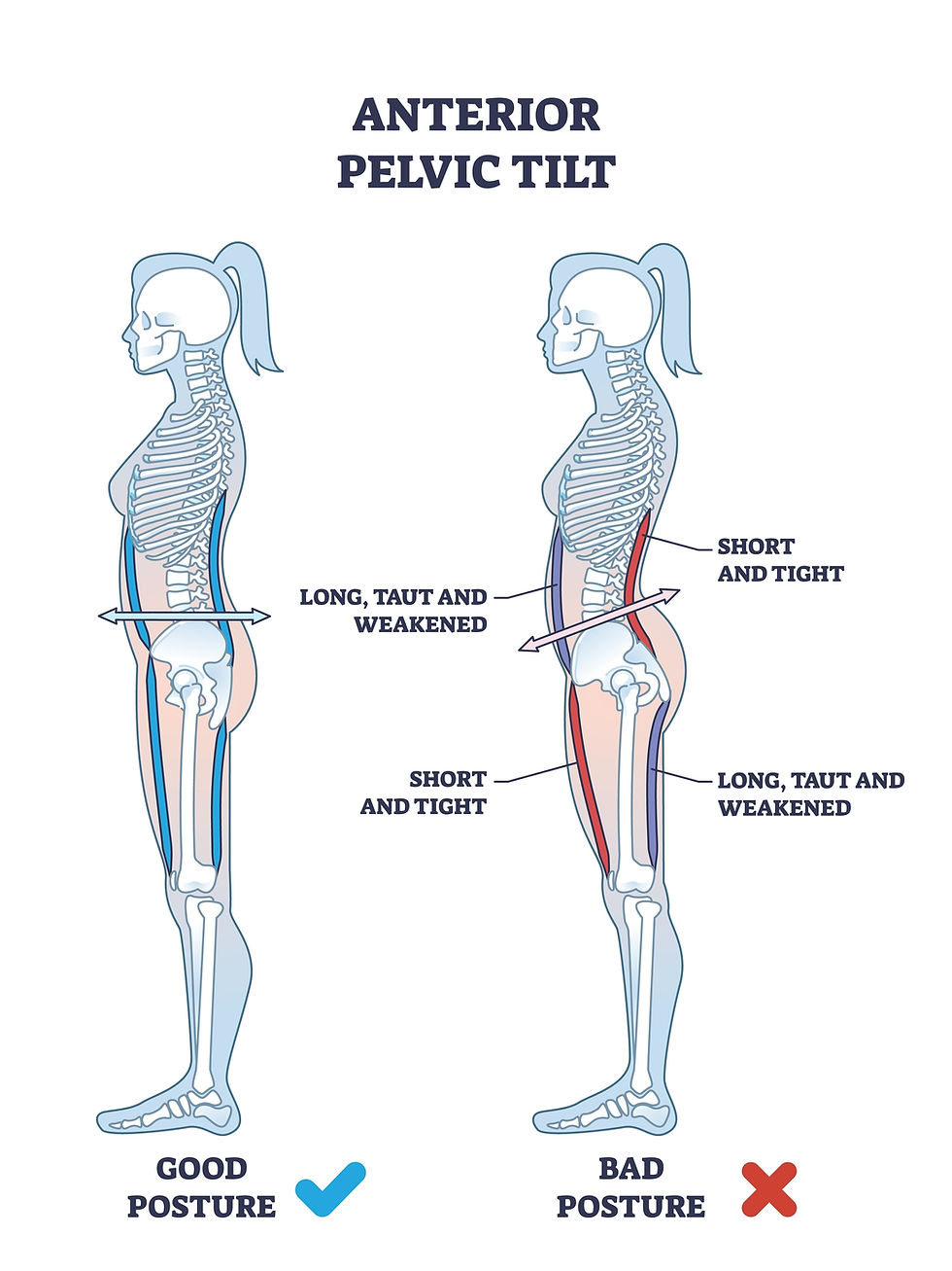
There is a correlation with both feet having increased pronation and developing an anterior pelvic tilt. Overpronation can be secondary to a lack of dorsiflexion ROM. An anterior pelvic tilt increases the risk of hamstring injury and pelvic floor dysfunction.
Proper dorsiflexion maintains stability and balance during walking and running. With a stable platform, you can push off the ground with greater force. Energy transfer is more effective when you have an optimal foot strike. Optimal dorsiflexion allows the effective engagement of the lower leg muscles and provides greater forward momentum during running.

Many areas of the brain light up every time your foot touches the ground. Not only does proper dorsiflexion impact sports performance in something like soccer, but it has even been shown to affect the way a volleyball player vertically swings their arm!!!
Dorsiflexion is correlated with swimming time, hand-grip strength, weightlifting performance, surfing performance, and, of course, soccer performance. Lower leg strength is increased with proper dorsiflexion.

Shoes and Dorsiflexion
Shoes make a huge difference in how the foot moves. As an example, in a study that was measuring foot pronation, the same person showed ankle joint eversion measuring at 31 degrees in one pair of shoes and 12 degrees in another pair. Studies on dorsiflexion have shown that dorsiflexion ROM is significantly greater in barefoot runners. Rigid shoes, shoes with a high heel-to-toe drop, or shoes with excessive cushioning can hinder dorsiflexion. With this in mind, I suggest researching shoe options. Zero-drop shoes offer a shoe with no elevation change between the heel and toe and promote natural foot positions. Still, you may need to transition into this type of shoe while performing activities to strengthen and support your feet.
There is also the thought that many of us need a shoe with a stiff heel, as that can stimulate a nerve on the bottom of the foot. Stimulating that nerve relaxes the hip flexors. SO if you are in zero-drop shoes and have tight hip flexors, one of the things you can consider is moving to a shoe with a supportive or stiffer heel. In this way, you are noticing what works for you. And many things create tight hip flexors, not just a nerve on the bottom of the foot. I regularly stand on a spiky mat, stimulate the nerves, and walk barefoot outdoors. I also wear zero-drop shoes.
The best advice on shoes is that you do not get stuck in a single pair. The foot needs different surfaces to stay happy. More posts on this and nerve stimulation to come!
Head to my shop page HERE to see what shoes and even socks I prefer.
How to Asses your Level of Dorsiflexion
Many types of practitioners can assess ankle dorsiflexion—such as chiropractors, PTs and manual therapists. And please know that within these fields, knowledge varies. I feel like a PT office would offer exact measurements of dorsiflexion. A manual therapist may not offer measurements, but if they work with a lot of athletes and ankle mobility, direct on-the table testing and therapy will illuminate the issue. And chiropractors (some more than others in my experience) offer ankle mobility testing and direct joint therapy.
But again, if you have recurring knee or hip issues, you can check in—on your own—with dorsiflexion. And of course, there is always You Tube and people on that platform will be able to demonstrate dorsiflexion for you! But you can also read the signs of ankle dysfunction, such as tight calves, and consider your own symptoms.
Knee to Wall Test
When doing a knee-to-wall test, a very general number is about 5 inches (12.5 cm) of space between your foot and the wall.
For this test, line yourself up facing a wall. Standing, feet hip distance apart. If you do not feel flexible, you can start with a small amount of space between your foot and the wall. Working your way up to testing your dorsiflexion at the 5 inches mark.
Keep your feet and heel planted firmly on the ground. Contact all four corners of your feet. As you lunge, make sure your knee stays centered, moving straight forward. If you see your knee moving inwards, then you are compensating. Stop and move yourself closer to the wall.
You can use a ruler between the wall and your toes to measure how far away you are from the wall. And you can measure your improvement over time.
_______________________________________________________________________________________________________________
Want to Know More?
I have more posts coming soon,. Stay tuned!
Check out my books on health and wellness!

The Elemental Woman
A Conversation for the Modern Western Woman Inspired by the Healing Wisdom of the Ancient Eastern Sage

Food & Mood Journal
A tracking guide to connect the food you eat, the emotions you feel, and increase the feeling of deep self-love

Mindful Movement Journal
A tracking guide to connect the motions you make, the emotions you feel, and increase the feeling of deep self-love

Basic Ayurveda
The Elemental Woman Supplement Guide
About the author:
Since 2004, Tara Lee Clasen has been assisting women on their healing adventures. As a woman-focused physical medicine provider, also trained in Eastern medicine, she knows transformation is possible and knows that with reflection and self-love, your future is full of bright possibilities.
#tmjpain #tmj #neckpain #shoulderpain #ribpain #chronicpain #jawpain #headaches #migraine #whiplash #asthma #vagusnerve #massagetherapy #manualtherapy #chiropratic #tmjpain #tmj #neckpain #shoulderpain #ribpain #chronicpain #jawpain #headaches #migraine #whiplash #asthma #vagusnerve #massagetherapy #manualtherapy #chiropractic #feet #Ankle #lowerleg #kneepain #ACL #ACLtear #meniscus #anklepain #dorsiflex #plantarflex #Anklemovement
Resources:
Hintermann B, Nigg BM (September 1998). "Pronation in runners. Implications for injuries". Sports Med. 26 (3): 169–76. doi:10.2165/00007256-199826030-00003. PMID 9802173. S2CID 24812917.
Martinez AF, Scattone Silva R, Paschoal BLF, Souza LLA,
Serrão FV. Association of ankle dorsiflexion and landing
forces in jumping athletes. Sports Health. 2022;14(6):932-7.
34961379 PMCid:PMC9631040
Fong CM, Blackburn JT, Norcross MF, McGrath M, Padua DA.
Ankle-dorsiflexion range of motion and landing
biomechanics. J Athl Train. 2011;46(1):5-10. https://doi.org
/10.4085/1062-6050-46.1.5 PMid:21214345 PMCid:
PMC3017488
Demirkan E, Ozkadi T, Can S, Alagoz I. Does ankle plantar
and dorsiflexion affect fifty-meter swimming time in
swimmers? Turk J Sport Exerc. 2021;23(3):353-8.
Abdulmassih S, Phisitkul P, Femino JE, Amendola A. Triceps
surae contracture: Implications for foot and ankle surgery.
J Am Acad Orthop Surg. 2013;21(7):398-407. https://doi.org
/10.5435/JAAOS-21-07-398 PMid:23818027
Teyhen DS, Shaffer SW, Butler RJ, et al. What risk factors are
associated with musculoskeletal injury in US army rangers?
A prospective prognostic study. Clin Orthop Relat Res.
2015;473(9):2948-58. https://doi.org/10.1007/s11999-015-
4342-6 PMid:26013150 PMCid:PMC4523518
Teyhen DS, Shaffer SW, Butler RJ, et al. What risk factors are
associated with musculoskeletal injury in US army rangers?
A prospective prognostic study. Clin Orthop Relat Res.
2015;473(9):2948-58. https://doi.org/10.1007/s11999-015-
4342-6 PMid:26013150 PMCid:PMC4523518
Chon SC, Chang KY, You JS. Effect of the abdominal draw-
in manoeuvre in combination with ankle dorsiflexion in
strengthening the transverse abdominal muscle in healthy
young adults: A preliminary, randomized, controlled study.
Physiotherapy. 2010;96(2):130-6. https://doi.org/10.1016/
j.physio.2009.09.007 PMid:20420959
Brantingham JW, Lee Gilbert J, Shaik J, Globe G. Sagittal
plane blockage of the foot, ankle and hallux and foot
alignment-prevalence and association with low back pain.
J Chiropr Med. 2006;5(4):123-7. https://doi.org/10.1016/
S0899-3467(07)60144-X PMid:19674683
Secomb JL, Farley ORL, Lundgren L, et al. Associations
between the performance of scoring manoeuvres and
lower-body strength and power in elite surfers. Int J Sports
Sci Coach. 2015;10(5):911-8. https://doi.org/10.1260/1747-
9541.10.5.911
Dowse RA, Secomb JL, Bruton M, Nimphius S. Ankle
proprioception, range of motion and drop landing ability
differentiates competitive and non-competitive surfers. J
Sci Med Sport. 2021;24(6):609-13. https://doi.org/10.1016/
j.jsams.2020.12.011 PMid:33414023
Francia P, Toni S, Iannone G, et al. How ankle joint mobility
changes in young soccer players of different ages: A time
series analysis. J Phys Educ Sport. 2021;21:2173-82.
Didnt use but intersting, brain
auber B, Gollhofer A, Taube W. Differences in motor
cortical control of the soleus and tibialis anterior. J Exp
Biol. 2018;221(Pt 20):jeb174680. https://doi.org/10.1242/
jeb.174680 PMid:30194250
Brain
Jiang T, Wu W, Wang X, Weng C, Wang Q, Guo Y. Activation
of brain areas following ankle dorsiflexion versus plantar
flexion: Functional magnetic resonance imaging
verification. Neural Regen Res. 2012;7(7):501-5.
Francis S, Lin X, Aboushoushah S, et al. fMRI analysis of
active, passive and electrically stimulated ankle
dorsiflexion. Neuroimage. 2009;44(2):469-79.
18950717
Trinastic JP, Kautz SA, McGregor K, et al. An fMRI study of
the differences in brain activity during active ankle
dorsiflexion and plantarflexion. Brain Imaging Behav.
2010;4(2):121-31. https://doi.org/10.1007/s11682-010-
9091-2 PMid:20502995
Papaiakovou G. Kinematic and kinetic differences in the
execution of vertical jumps between people with good and
poor ankle joint dorsiflexion. J Sports Sci. 2013;31(16):
PMid:23879544
Stanley LE, Harkey M, Luc-Harkey B, et al. Ankle
Dorsiflexion displacement is associated with hip and knee
kinematics in females following anterior cruciate ligament
reconstruction. Res Sports Med. 2019;27(1):21-33.
30084269
Rabin A, Portnoy S, Kozol Z. The association of ankle
dorsiflexion range of motion with hip and knee kinematics
during the lateral step-down test. J Orthop Sports Phys
Ther. 2016;46(11):1002-9. https://doi.org/10.2519/jospt.
2016.6621 PMid:27686412
Panoutsakopoulos V, Kotzamanidou MC, Papaiakovou G,
Kollias IA. The ankle joint range of motion and its effect on
squat jump performance with and without arm swing in
adolescent female volleyball players. J Funct Morphol
Kinesiol. 2021;6(1):14. https://doi.org/10.3390/jfmk
6010014 PMid:33546291 PMCid:PMC7931004
Hoch MC, Staton GS, McKeon PO. Dorsiflexion range of
motion significantly influences dynamic balance. J Sci Med
Sport. 2011;14(1):90-2. https://doi.org/10.1016/j.jsams.
2010.08.001 PMid:20843744
Howe LP. The acute effects of ankle mobilizations on lower
extremity joint kinematics. J Bodyw Mov Ther.
2017;21(4):775-80. https://doi.org/10.1016/j.jbmt.2016.11.
007 PMid:29037626
Macrum E, Bell DR, Boling M, Lewek M, Padua D. Effect of
limiting ankle-dorsiflexion range of motion on lower
extremity kinematics and muscle-activation patterns
during a squat. J Sport Rehabil. 2012;21(2):144-50.
https://doi.org/10.1123/jsr.21.2.144 PMid:22100617
Witvrouw E, Lysens R, Bellemans J, Cambier D,
Vanderstraeten G. Intrinsic risk factors for the development
of anterior knee pain in an athletic population. A two-year
prospective study. Am J Sports Med. 2000;28(4):480-9.
10921638
Piva SR, Goodnite EA, Childs JD. Strength around the hip
and flexibility of soft tissues in individuals with and without
patellofemoral pain syndrome. J Orthop Sports Phys Ther.
2005;35(12):793-801. https://doi.org/10.2519/jospt.2005.
35.12.793 PMid:16848100
Malloy P, Morgan A, Meinerz C, Geiser C, Kipp K. The
association of dorsiflexion flexibility on knee kinematics
and kinetics during a drop vertical jump in healthy female
athletes. Knee Surg Sports Traumatol Arthrosc. 2015;
23(12):3550-5. https://doi.org/10.1007/s00167-014-3222-z
PMid:25112598 PMCid:PMC4977993
Noehren B, Hamill J, Davis I. Prospective evidence for a hip
etiology in patellofemoral pain. Med Sci Sports Exerc.
2013;45(6):1120-4. https://doi.org/10.1249/MSS.0b013e
31828249d2 PMid:23274607
Greiwe RM, Saifi C, Ahmad CS, Gardner TR. Anatomy and
biomechanics of patellar instability. Oper Tech Sports Med.
2010;18(2):62-7. https://doi.org/10.1053/j.otsm.2009.12.
014
Boden BP, Sheehan FT, Torg JS, Hewett TE. Noncontact
anterior cruciate ligament injuries: Mechanisms and risk
factors. J Am Acad Orthop Surg. 2010;18(9):520-7.
20810933 PMCid:PMC3625971
Aali S, Rezazadeh F, Badicu G, Grosz WR. Effect of heel-first
strike gait on knee and ankle mechanics. Medicina
(Kaunas). 2021;57(7):657. https://doi.org/10.3390/
medicina57070657 PMid:34206943 PMCid:PMC8304808
Kuhman DJ, Paquette MR, Peel SA, Melcher DA.
Comparison of ankle kinematics and ground reaction
forces between prospectively injured and uninjured
collegiate cross country runners. Hum Mov Sci. 2016;47:9-
26827155
Koga H, Muneta T, Bahr R, Engebretsen L, Krosshaug T. ACL
injury mechanisms: Lessons learned from video analysis.
In: Musahl V, Karlsson J, Kuroda R, Zaffagnini S, editors.
Rotatory knee instability. Springer; 2017. p. 27-36.
Leppänen M, Pasanen K, Kujala UM, et al. Stiff landings are
associated with increased ACL injury risk in young female
basketball and floorball players. Am J Sports Med.
2017;45(2):386-93. https://doi.org/10.1177/036354651666
5810 PMid:27637264
Waldén M, Krosshaug T, Bjørneboe J, Andersen TE, Faul O,
Hägglund M. Three distinct mechanisms predominate in
non-contact anterior cruciate ligament injuries in male
professional football players: A systematic video analysis of
39 cases. Br J Sports Med. 2015;49(22):1452-60.
25907183 PMCid:PMC4680158
Souza TR, Pinto RZ, Trede RG, Kirkwood RN, Pertence AE,
Fonseca ST. Late rearfoot eversion and lower-limb internal
rotation caused by changes in the interaction between
forefoot and support surface. J Am Podiatr Med Assoc.
2009;99(6):503-11. https://doi.org/10.7547/0990503 PMid:
19917736
Mendonça LD, Verhagen E, Bittencourt NF, Gonçalves GG,
Ocarino JM, Fonseca ST. Factors associated with the
presence of patellar tendon abnormalities in male athletes.
J Sci Med Sport. 2016;19(5):389-94. https://doi.org/10.
1016/j.jsams.2015.05.011 PMid:26087883
Yu B, Garrett WE. Mechanisms of non-contact ACL injuries.
Br J Sports Med. 2007;41 Suppl 1(Suppl 1):i47-51.
https://doi.org/10.1136/bjsm.2007.037192 PMid:17646249
PMCid:PMC2465243
Baumbach SF, Braunstein M, Seeliger F, Borgmann L,
Böcker W, Polzer H. Ankle dorsiflexion: What is normal?
Development of a decision pathway for diagnosing
impaired ankle dorsiflexion and M. gastrocnemius
tightness. Arch Orthop Trauma Surg. 2016;136(9):1203-11.
https://doi.org/10.1007/s00402-016-2513-x PMid:27418341
Malliaras P, Cook JL, Kent P. Reduced ankle dorsiflexion
range may increase the risk of patellar tendon injury among
volleyball players. J Sci Med Sport. 2006;9(4):304-9.
16672192
Hamilton M, Velasquez JR. Ankle flexibility and jump
landing mechanics: Implications for ACL injury risk. Int J
Athl Ther Train. 2011;16(6):14-6. https://doi.org/10.1123/
ijatt.16.6.14
Pascual Huerta J. The effect of the gastrocnemius on the
plantar fascia. Foot Ankle Clin. 2014;19(4):701-18.
https://doi.org/10.1016/j.fcl.2014.08.011 PMid:25456717
DiGiovanni CW, Langer P. The role of isolated
gastrocnemius and combined Achilles contractures in the
flatfoot. Foot Ankle Clin. 2007;12(2):363-79, viii.
https://doi.org/10.1016/j.fcl.2007.03.005 PMid:17561207
Mahieu NN, Witvrouw E, Stevens V, Van Tiggelen D, Roget P.
Intrinsic risk factors for the development of achilles tendon
overuse injury: A prospective study. Am J Sports Med. 2006;
34(2):226-35. https://doi.org/10.1177/0363546505279918
PMid:16260469
Pinto RZ, Souza TR, Trede RG, Kirkwood RN, Figueiredo EM,
Fonseca ST. Bilateral and unilateral increases in calcaneal
eversion affect pelvic alignment in standing position. Man
Ther 2008;13(6):513-9. https://doi.org/10.1016/j.math.
2007.06.004 PMid:17910932
Alizadeh S, Mattes K. How anterior pelvic tilt affects the
lower extremity kinematics during the late swing phase in
soccer players while running: A time series analysis. Hum
Mov Sci. 2019;66:459-66. https://doi.org/10.1016/j.humov.
2019.06.001 PMid:31176257
PF
Kannan P, Winser S, Goonetilleke R, Cheing G. Ankle
positions potentially facilitating greater maximal
contraction of pelvic floor muscles: A systematic review
and meta-analysis. Disabil Rehabil. 2019;41(21):2483-91.
29733699
Aquino MRC, Resende RA, Kirkwood RN, Souza TR, Fonseca
ST, Ocarino JM. Spatial-temporal parameters, pelvic and
lower limb movements during gait in individuals with
reduced passive ankle dorsiflexion. Gait Posture.
2022;93:32-8. https://doi.org/10.1016/j.gaitpost.2022.01.
010 PMid:35063755


Comments